Introduction
Chest drainage tubes, also known as thoracostomy tubes or simply “chest tubes,” are vital medical devices used to remove air, fluids, or pus from the pleural space, restoring proper lung function and relieving respiratory distress. These tubes are often a critical component in treating conditions such as pleural effusion, pneumothorax, and heart failure. This comprehensive guide will delve into the intricacies of chest drainage tubes, from their placement in the chest cavity to the meticulous care they require.
Understanding the Pleural Space
The pleural space is a slender, fluid-filled gap between the lung’s visceral pleura and the chest wall’s parietal pleura. This space allows for smooth lung movement during respiration. However, when excess fluid, blood, or air accumulates in the pleural cavity, it can lead to a collapsed lung or pleural effusion, causing significant respiratory distress and requiring immediate medical intervention.
Types of Chest Drainage Tubes
When someone refers to chest tubes, you have to consider the clinical context. There are several types of chest drainage tubes, each designed for specific conditions:
- Small-bore chest tubes: Often used for less severe pleural effusions or pneumothorax. They are less invasive and can be placed using the Seldinger technique.
- Large-bore chest tubes are required for large volumes of fluid or air, such as in cases of trauma or after chest surgery. They are normally placed using a bedside procedure or during surgery.
- Pigtail catheters: Flexible tubes typically used for draining pleural fluid in a less invasive manner. These are often placed by interventional radiology.
- Tunneled pleural catheters: For long-term use, patients with chronic effusions can manage their condition at home.
Chest Tube Placement Procedure
Chest tube insertion, or tube thoracostomy, is typically performed under local anesthesia. However, general anesthesia may be used when chest tubes are placed as part of a thoracic surgery. The steps include:
- Administering local anesthetic at the incision site.
- Making a small incision in the chest wall, often near the inframammary fold for cosmetic reasons.
- Carefully creating a path through the subcutaneous tissue and intercostal muscles.
- Inserting the chest tube into the pleural space over the rib, and ensuring the number of drainage holes are within the chest cavity.
- The tube is connected to a chest tube drainage system, which often includes a water seal chamber to prevent air re-entry and a collection chamber for the fluid.
Chest Drainage Tube Care and Monitoring
After placement of a chest tube, it is essential to:
- Monitor the amount of fluid and check for any sudden increase in drainage, which could indicate bleeding.
- Ensure the water level in the water seal chamber fluctuates with breathing, indicating the system is connected and functioning.
- Regularly assess the patient’s vital signs, including heart rate and blood pressure.
- Provide regular pain relief to manage discomfort at the tu
- be insertion site.
Patients will undergo frequent chest radiographs to confirm the right place of the tube and the re-expansion of the lung.
Complications of Chest Drainage Tubes
While chest tube insertion is a life-saving procedure, it comes with potential risks such as:
- Infection: Signs of infection at the incision site must be monitored by the medical team.
- Air leak: Persistent air leaks suggest a continued breach in the lung or pleural space.
- Blood clot: A clot may obstruct the tube, necessitating prompt medical attention.
Chest Tube Removal
Removal of the chest tube is considered when:
- The underlying condition has resolved, evidenced by a lack of air leak and minimal fluid drainage.
- The chest x-ray shows complete lung expansion.
- The patient can breathe comfortably and maintain oxygen levels without assistance.
The healthcare provider will remove the tube, often after a trial of clamping to ensure the lung remains expanded without the tube in place.
The Role of the Health Care Provider
The health care provider plays a crucial role in the management of chest drainage systems. They are responsible for assessing the patient’s thoracic cavity, determining the need for a thoracostomy tube, and ensuring that the negative pressure within the chest tube drainage system is appropriately maintained. This negative pressure is vital for the re-expansion of the lung and the removal of excess air and fluid. Providers must also instruct patients on how to take deep breaths effectively, which can help re-expand the lung and clear any pleural fluid.
The Mechanics of Chest Drainage Systems
Chest drainage systems typically consist of a plastic tube that is inserted through the chest wall into the pleural space. This intercostal drain allows for the removal of air and fluid from the thoracic cavity, often using a wet suction system that includes a suction control chamber filled with sterile water. The amount of suction is carefully regulated by the healthcare professional to ensure the patient’s comfort and the effective drainage of the chest cavity.
Patient Comfort and Monitoring
Pain management is a critical aspect of patient care post-chest tube insertion. Pain medication is administered to manage the discomfort associated with the procedure and the presence of the tube. Additionally, the patient is monitored using a chest radiograph to confirm the location of the chest tube and to assess the lung’s re-expansion. A sterile dressing is applied to the site of insertion to minimize the risk of infection and to promote healing, leaving only a small scar once the tube is removed.
Advanced Chest Drainage Technology
Modern chest drainage systems have evolved to include safety features that minimize the risk of complications. For instance, flexible tubes are now commonly used, which are less rigid and more comfortable for the patient. Wet suction systems have been supplemented with dry suction and water seal systems, which are easier to manage and more mobile. These systems often include a one-way valve, which prevents backflow of air or fluid into the patient, a critical feature in the management of conditions like tension pneumothorax.
Surgical Considerations and Complications
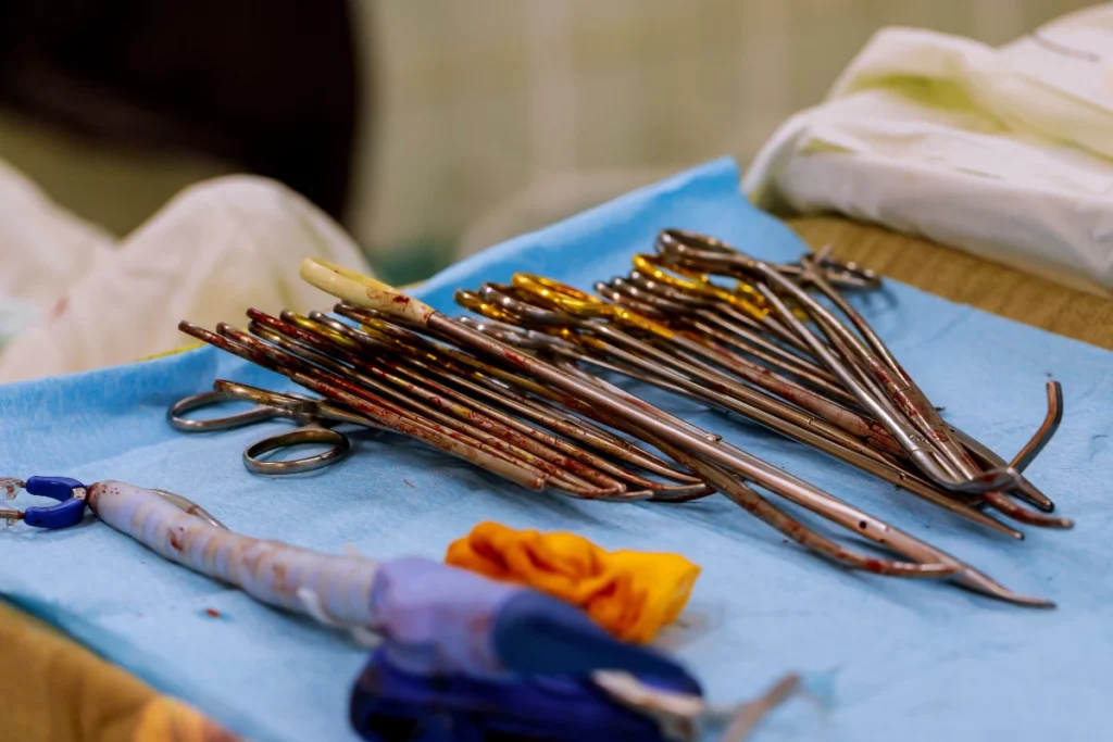
In the operating room, the placement of a chest tube is often the end of a surgical procedure, especially in lung surgery or after interventions that may compromise respiratory function. The procedure, while invasive, is necessary to prevent serious complications such as subcutaneous emphysema, where air accumulates under the skin, or a tension pneumothorax, which can be life-threatening. Sterile drapes are used to maintain a clean environment, and a local anaesthetic is administered at the site of insertion to reduce pain.
Long-Term Management and Removal
For patients requiring long-term drainage, such as those with chronic pleural effusions or on mechanical ventilation, tunneled pleural drains or indwelling pleural catheters may be used. These allow for the management of pleural fluid in an outpatient setting. The removal of a chest tube is a significant step indicating the patient’s recovery. It requires careful monitoring by the healthcare professional to ensure the lung remains expanded and that there is no recurrence of fluid accumulation.
Conclusion
Chest drainage tubes play a pivotal role in managing pleural space conditions, from emergency department thoracostomies to planned thoracic surgeries. Their success is dependent on careful monitoring, understanding the mechanics of the closed chest drainage system, and addressing complications promptly.
Chest drainage tubes are a cornerstone in the management of various thoracic conditions. From the traditional chest drainage system to the latest in thoracostomy technology, the goal remains the same: to alleviate difficulty breathing and restore normal respiratory function. The healthcare team, including the healthcare provider and the healthcare professional, must work in tandem to ensure the safe and effective use of these devices, minimizing the risk of complications and ensuring the best possible outcomes for the patient.
Additional Considerations
FAQs about Chest Drainage Tubes
Here are some common questions and answers to help demystify the use of chest drainage tubes:
- Q: How long does a chest tube stay in place?
- A: It varies depending on the condition being treated and the patient’s response to therapy.
- Q: Is the insertion procedure painful?
- A: Local anesthesia and sedation are used to minimize discomfort.
- Q: Can I shower with a chest tube?
- A: Healthcare professionals will provide specific instructions on showering and dressing.
For further reading and understanding, consider these resources:

